Common Name of Agent/Disease: White spot syndrome virus (WSSV), white spot syndrome (WSS) (Durand et al., 1996; Lightner, 1996; Lo et al., 1996a).
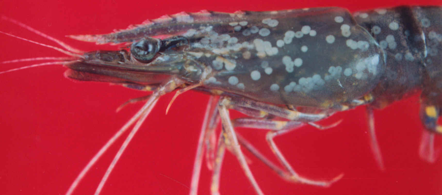
Fig. Tiger Shrimp (P. monodon) showing typical white spots. Photo.: A. Gopalakrishnan
Distinguishing Features: White spot syndrome virus (WSSV) is
rod-shaped, enveloped, non-occluded virions. The nucleocapsid of the virion is
composed of subunits in ring formation that are stacked in a series and almost
prependicular to the longitudinal axis of the capsid. Apparent in some virions
is a tail-like projection extending from one end which is an envelope extension
(Wang et al., 1995; Durand et al., 1997). The nucleocapsid is closed and rounded
at one end and squared at the other (Durand et al., 1997). Reports on the size
of the virion vary due to different methods of preparing the samples and
different measurement techniques employed. In addition, Kasornchandra et al.
(1998) found that viral isolates from different geographic areas, when prepared
similarly, do have intrinsic size variations. The complete virions range in size
from 83-120 nm X 270-330 nm (Kasornchandra et al., 1998). The smallest was the
sample from Japan and the largest was from China. The virion contains
double-stranded DNA that is 190-200 kilobases in length. There are three major
proteins composing WSBV with molecular weights of 19, 23.5, and 27.5 kDa (Cesar
et al., 1998).
Similar Species: Due to the morphology, size, site of assembly,
cellular pathology, and nucleic acid of white spot syndrome virus (WSSV) it was
originally proposed to be placed in the genus Non-Occluded Baculovirus,
subfamily Nudibaculovirinae, and family Baculoviridae. One other
virus of penaeid shrimp, the baculoviral midgut gland necrosis virus (BMNV), was
a proposed member of this subfamily at that time. In 1995, the International
Committee on Taxonomy of Viruses (ICTV) deleted the genus Non-Occluded
Baculovirus and the subfamily Nudibaculovirinae and left the viruses
previously in this classification as unassigned invertebrate viruses (Murphy et
al. 1995). Other members of this previous classification include two insect
viruses, Oryctes rhinoceros virus and Heliothis zea virus 1. For consistency,
many authors have maintained the old designation as a baculovirus and use the
name white spot syndrome baculovirus or more commonly white spot syndrome virus.
Two viruses of crabs are similar to white spot syndrome virus in form and location of replication. These include the baculovirus of the crab Carcinus maenas, and B2 in C. mediterraneus (Durand et al., 1996).
Biology: Taxonomic and Nomenclature Remarks: The virus has been found at multiple geographic locations giving rise to several names for this disease. These names have been derived from the appearance of the animals infected or to the species or tissue types where it has been found. For example, in Japan, Inouye et al. (1994) named the virus they discovered rod-shaped nuclear virus of Penaeus japonicus (RV-PJ). This is based upon the host species and the shape of the virus when viewed by transmission electron microscopy. Woongteerasupaya et al. (1995) named the virus found in Thailand systemic ectodermal and mesodermal baculovirus (SEMBV). This virus infected circulating hemocytes (therefore systemic), cells of ectodermal and mesodermal origin, and the virion (single virus particle) itself resembled those of the family Baculoviridae. Chou et al. (1995) of Taiwan named the virus white spot syndrome. This name refers to the obvious white spots on the carapace, appendages, and the inside surface of the body of the infected shrimp. It has generally been accepted that all of these isolates are strains of the same entity and the name of white spot syndrome virus is utilized universally.
To which family to assign this virus remains uncertain. Following the guidelines set forth by the fifth report of the International Commitee on Taxonomy of Viruses (ICTV) (Francki et al., 1991) Wang et al. (1995) suggested that this virus is a member of the genus Non-Occluded Baculovirus, the subfamily Nudibaculovirinae, and the family Baculoviridae. Later, in 1995, the sixth report of the ICTV canceled the classification of Nudibaculovirinae and Non-Occluded Baculovirus (Murphy et al., 1995). The viruses previously classified as members of this genus and subfamily, such as the insect viruses Oryctes rhinoceros virus and Heliothis zea virus 1, are now listed as unassigned invertebrate viruses. For consistency the name of white spot syndrome virus has been maintained.
Morphology: The white spot syndrome virus (WSSV) is a rod-shaped, enveloped virus with an envelope extension from one end that appears to be a tail (Wang et al., 1995; Durand et al., 1997). The capsid is composed of rings of subunits in a stacked array. The rings are perpendicular to the longitudinal axis of the capsid (Wang et al., 1995; Durand et al., 1997; Cesar et al., 1998). One end of this cylindar of subunits is closed by a rounded segment. The other end is squared (Durand et al., 1997). The virus contains double stranded DNA that is approximately 190-200 kilobases in length. Three major proteins compose this virus and they have molecular weights of 19, 23.5, and 27.5 kDa (Cesar et al., 1998).
Pathology: Shrimp that are acutely affected by white spot syndrome virus (WSSV) show a rapid reduction in food consumption, lethargy, a loose cuticle, and a pink to reddish-brown discoloration of the hepatopancreas (Chou et al., 1995; Lightner, 1996). White spots form on the inside of the carapace after or at the end of the acute phase of infection in some animals. There is some debate over the presence of white spots in American penaeids (Lightner and Lotz, personal communication). The white spots are 0.5 to 2.0 mm in diameter and are caused by abnormal deposits of calcium salts by the cuticular epidermis (Lightner, 1996). This virus can cause mortalities of 100% within 3 to 10 days of the onset of the above symptoms (Lightner, 1996). WSSV has also been found to exist asymptomatically (without symptoms) in wild shrimp and crabs for extended periods of time. It only causes symptoms and mortalities when the animals are placed under stress, such as in captivity or when spawned (Lo et al., 1996b, 1997).
Diagnostic techniques:
Gross Observations: Acutely infected shrimp often have a loose cutical with white spots (which represent abnormal deposits of calcium salts by the cuticular epidermis) of 0.5 - 2.0 mm in diameter which are most apparent on the inside surface of the carapace. In many cases morbund shrimp displayed a pink to reddish-brown colouration due to expansion of the cuticular chromatophores and few if any white spots.
Squash Preparations: Hypertrophied or vacuolated nuclei usually with a single eosinophilic to bluish inclusion body in squashes or impression smears (stained with Giemsa or other blood smear stains) of epithelia and connective tissues of the gills or stomach of shrimp with clinical signs. Occlusion bodies are absent. Normal cell nuclei are 4 - 10 µm in diameter and display chromatin threads and a nucleolus.
Histology: Hematoxylin and eosin staining of white spot syndrome virus infected tissue presents eosinophilic inclusion bodies with hypertrophied (swollen) nuclei and marginated, slightly basophilic, chromatin. The nuclear hypertrophy is due to the development and accumulation of developing virions within the nucleus. The cuticular epithelial cells and connective tissue cells, and, less frequently, the antenal gland epithelium, lymphoid organ sheath cells, haematopoietic cells and fixed phagocytes of the heart are the target cells. Occlusion bodies are absent. In the early stages of inclusion body development, they are eosinophilic, centronuclear, with a halo (an artifact with Davidson's fixation) and resemble the inclusion bodies of IHHNV. However, the presence of larger more fully developed (without a halo) pale basophilic inclusion bodies in infected target tissue cells during the advanced stages of infection clearly distinguishes the two diseases.
In later stages of development, the inclusions become more basophilic in appearance (Durand et al., 1996, 1997; Lightner, 1996; Wongteerasypaya et al., 1995). WSSV targets predominately cuticular epithelium and subsequently connective tissues of some organs. The virus severely damages the stomach, gills, subcuticular epithelial cells, lymphoid organ, antennal gland and hemocytes. Also prone to infection, but less frequently, are the antennal gland epithelium, lymphoid organ sheath cells, nerve node, hepatocpancreas, and striated muscle (Chang et al.,1996; Lightner, 1996).
Electron Microscopy: Cytopathology occurs in the appropriate target tissue types and is accompanied by large rod-shaped to somewhat elliptical, non-occluded virions of about 70 - 150 nm in width and about 275 - 380 nm in length in the intranuclear inclusion bodies of infected cells.
DNA Probes: WSBV infected nuclei can be intensely marked by a DIG-labeled DNA probe for WSBV with in situ hybridization assays. Gene probes for WSBV are being developed in China, Japan, Thailand and at the University of Arizona in the U.S. None of the WSBV complex are reactive to the available gene probes to IHHNV, BP, MBV and HPV.
Host Species Affected by the Virus: Forty-three species are known to be hosts of the white spot syndrome virus world wide. Penaeus monodon, Penaeus japonicus, Penaeus chinensis (=orientalis), Penaeus indicus, Penaeus merguiensis, Penaeus penicillatus and Penaeus setiferus (in Texas). Experimentally, severe and lethal infections of WSBV from Thailand were produced in Penaeus vannamei, Penaeus stylirostris, Penaeus aztecus, Penaeus duorarum and Penaeus setiferus. No significant resistance to WSBV complex has been reported for any of the penaeid shrimp. In the Gulf of Mexico the following species are potential hosts of WSSV: Litopenaeus setiferus, Farfantepenaeus duorarum, and F. aztecus.
Distribution: White spot disease was first detected in shrimp farms in Texas in 1995. Strains of WSSV are wide spread throughout China, Japan, Korea and Thailand (Lightner, 1996).
Interest to Fisheries: White spot syndrome virus is a serious threat to the shrimp industry. WSSV infects a very broad spectrum of crustaceans. Some species do not succumb to the disease, but are carriers able to spread the pathogen. It is very important to eliminate these carrier species from shrimp farming systems (Flegel, 1996).
Current Status of this Species in the Gulf of Mexico Ecosystem: White spot syndrome virus was found in coastal Texas culturing facilities in 1995 (Lightner, 1996).
Potential Impacts:
Assessment of Risks: White spot syndrome virus (WSSV) causes cumulative mortalities of up to 100% within 3 to 10 days of the first signs of the disease (Lightner, 1996). Dr. D.V. Lightner of the University of Arizona estimates that since its discovery in 1992, WSSV is responsible for $4-6 Billion in cumulative loses (personal communication).
Nunan et al. (1998) have demonstrated that WSSV is present in some frozen imported shrimp products purchased from grocery stores. Frozen shrimp are utilized by sport fishermen as bait. This is a potential source of viral introduction into local, wild populations of shrimp (Humphrey, 1995). Also, shrimp are repackaged at processing plants in the United States and the solid waste, such as the carapace, are disposed of into local water or landfills (Overstreet et al., 1997). This is the implicated source of the first WSSV case in the Gulf of Mexico region in 1995 (Lightner, 1996).
Recommendations: The best approach to manage any viral disease is to implement preventative measures to keep it out of the production system. These include disinfecting ponds and eliminating potential viral carriers prior to stocking, the use of fine screens at water inlets to remove potential carriers, avoidance of fresh feed (not heat processed) products that may contain crustacean species that are carriers or hosts for WSSV, and disinfection of WSSV pond water before discharge (Flegel, 1996). Also, monitoring of all stocks through the use of stained squashes or impression smears of epithelial or connective tissue (Nunan and Lightner, 1997), histology (Lightner, 1996), in-situ hybridization (Nunan and Lightner, 1997), dot blot (Chang et al., 1998), or polymerase chain reaction (Lo et al., 1996a) for WSBV is an essential measure (Flegel, 1996).
References:
Chang, P.-S., C.-F. Lo, Y.-C. Wang, G.-H. Kou. 1996. Identification of white spot syndrome associated baculovirus (WSBV) target organs in the shrimp penaeus monodon by in situ hybridization. Diseases of Aquatic Organisms 27:131-139.
Chang, P.-S., D.-H. Tasi, Y.-C. Wang. 1998. Development and evaluation of a dot blot analysis for the detection of white spot syndrome baculovirus (WSBV) in Penaeus monodon.
Chou, H.-Y., C.-Y. Huang, C.-H. Wang, H.-C. Chiang, C.-F. Lo. 1995. Pathogenicity of a baculovirus infection causing white spot syndrome in cultured penaeid shrimp in Taiwan. Diseases of Aquatic Organisms 23:165-173.
Durand, S., D.V. Lightner, L.M. Nunan, R.M. Redman, J. Mari, J.-R. Bonami. 1996. Application of gene probes as diagnostic tools for White Spot Baculovirus (WSBV) for penaeid shrimp. Diseases of Aquatic Organisms 27:59-66.
Durand,S., D.V. Lightner, R.M. Redman, J.-R. Bonami. 1997. Ultrastructure and morphogenesis of whtie spot syndrome baculovirus (WSSV). Diseases of Aquatic Organisms 29:205-211.
Flegel, T.W. 1996. The white spot virus crisis in Asian shrimp culture. Aquaculture Asia July-September:29-34.
Francki, R.I.B., C.M. Fauquet, D.L. Knudson, F. Brown. 1991. Classification and nomenclature of viruses. Archives of Virology. Springer-Verlag, Vienna. Suppl 2:1-450.
Huang, J., X.L. Song, J. Yu, C.H. Yang. 1994. Baculoviral hypodermal and hematopoietic necrosis-pathology of the shrimp explosive epidemic disease. Yellow Sea Fishery Research Institute, Qingdao, PR, China (abstract).
Humphrey, J.D. 1995. Australian quarantine policies and practices for aquatic animals and their products: a review for the Scientific Working Party on Aquatic Animal Quarantine. Bureau of Resource Sciences, Cnaberra.
Inouye, K., S. Miwa, N. Oseko, H. Nakano, T. Kimura, K. Momoyama, and M. Hiraoka. 1994. Mass mortalities of cultured kuruma shrimp Penaeus japonicus in Japan in 1993: electron microscopic evidence of the causative virus. Fish Pathology 29(2):149-158.
Kanchanaphum, P., C. Woongteerasupaya, N. Sitidilokratana, V. Boonsaeng, S. Panyim, A. Tassanakajon, B. Withyachumnarnkul, T.W. Flegel. 1998. Experimental transmission of white spot syndrome virus (WSSV) from crabs to shrimp Penaeus monodon. Diseases of Aquatic Organisms 34:1-7.
Kasornchandra, J., S. Boonyaratpalin, T. Itami. 1998. Detection of whit-spot syndrome in cultured penaeid shrimp in Asia: microscopic observation and polymerase chain reaction. Aquaculture 164:243-251.
Lightner, D.V. 1996. A handbook of shrimp pathology and diagnostic procedures for diseases of cultured penaeid shrimp. World Aquaculture Society, Baton Rouge, Louisiana, USA.
Lo, C.-F., J.-H. Leu, C.-H. Ho, C.-H. Chen, S.-E. Peng, Y.-T. Chen, C.-M. Chou, P.-Y. Yeh, C.-J. Huang, H.-Y. Chou, C.-H. Wang, G.-H. Kou. 1996a. Detection of baculovirus associated with white spot syndrome (WSBV) in penaeid shrimps using polymerase chain reaction. Diseases of Aquatic Organisms 25:133-141.
Lo, C.-F., C.-H. Ho, S.-E. Peng, C.-H. Chen, H.-C. Hsu, Y.-L. Chiu, C.-F. Chang, K.-F. Liu, M.-S. Su, C.-H. Wang, G.-H. Kou. 1996b. White spot syndrome baculovirus (WSBV) detected in cultured and captured shrimp, crabs and other arthropods. Diseases of Aquatic Organisms 27:215-225.
Lo, C.-F., C.-H. Ho, C.-H. Chen, K.-F. Liu, Y.-L. Chiu, P.-Y. Yeh, S.-E. Peng, H.-C. Hsu, H.-C. Liu, C.-F. Chang, M.-S. Su, C.-H. Wang, G.-H. Kou. 1997. Detection and tissue tropism of white spot syndrome baculovirus (WSBV) in captured brooders of Penaeus monodon with a special emphasis on reproductive organs. Diseases of Aquatic Organisms 30:53-72.
Maeda, M., T. Itami, A. Furumoto, O. Hennig, T. Imamura, M. Kondo, I. Hirono, T. Aoki, Y. Takahashi. 1998. Detection of penaeid rod-shaped DNA virus (PRDV) in wild-caught shrimp and other crustaceans. Fish Pathology 33(4):373-380.
Murphy, F.A., C.M. Fauquet, D.H.L. Bishop, S.A. Ghabrial, A.W. Jarvis, G.P. Martelli, M.A. Mayo, M.D. Summers (eds.) 1995. Virus taxonomy. Sixth Report of the International Committee on Taxonomy of Viruses. Archives of Virology, Vienna: Springer, p.586.
Nadala Jr., E.C.B., P.C. Loh. 1998. A comparative study of three different isolates of white spot virus. Diseases of Aquatic Organisms 33:231-234.
Nunan, L.M., D.V. Lightner. 1997. Development of a non-radioacitve gene probe by PCR for detection of white spot syndrome virus (WSSV). Journal of Virological Methods 63:193-201.
Nunan, L.M., B.T. Poulos, D.V. Lightner. 1998. The detection of white spot syndrome virus (WSSV) and yellow head virus (YHV) in imported commodity shrimp. Aquaculture 160:19-30.
Overstreet, R.M., D.V. Lightner, d.W. Hasson, S. McIlwain, J.M. Lotz. 1997. Susceptibility to Taura syndrome virus of some penaeid shrimp species native to the Gulf of Mexico and the southeastern United States. Journal of Invertebrate Pathology 69:165-176.
Supamattaya, K., R.W. Hoffmann, S. Boonyaratpalin, P. Kanchanaphum. 1998. Experimental transmission of white spot syndrome virus (WSSV) from black tiger shrimp Penaeus monodon to the sand crab Portunus pelagicus, mud crab Scylla serrata and krill Acetes sp. Diseases of Aquatic Organisms 32:79-85.
Takahashi, Y., T. Itami, M. Kondo, M. Maeda, R. Fujii, S. Tomonaga, K. Supamattaya, S. Boonyaratpalin. 1994. Electron microscopic evidence of bacilliform virus infection in kuruma shrimp (Penaeus japonicus). Fish Pathology 29(2):121-125.
Wang, C.-H., C.-F. Lo, J.-H. Leu, C.-M. Chou, P.-Y. Yeh, H.-Y. Chou, M.-C. Tung, C.-F. Chang, M.-S. Su, G.-H. Kou. 1995. Purification and genomic analysis of baculovirus associated with white spot syndrome (WSBV) of Penaeus monodon. Diseases of Aquatic Organisms 23:239-242.
Wang, C.S., Y.J. Tsai, G.H. Kou, S.N. Chen. 1997. Detection of white spot disease virus infection in wild-caught greasy back shrimp, Metapenaeus ensis (de Haan) in Taiwan. Fish Pathology 32(1):35-41.
Wang, Q., B.L. White, R.M. Redman, D.V. Lightner. 1999. Per os challenge of Litopenaeus vannamei postlarvae and Farfantepenaeus duorarum juveniles with six geographic isolates of white spot syndrome virus. Aquaculture 170:179-194.
Wang, Y.-C., C.-F. Lo, P.-S. Chang, G.-H. Kou. 1998. Experimental infection of white spot baculovirus in some cultured and wild decapods in Taiwan. Aquaculture 164:221-231.
Wongteerasupaya, C., J.E. Vickers, S. Sriurairatana, G.L. Nash, A.
Akarajamorn, V. Boonsaeng, S. Panyim, A. Tassanakajon, B. Withyachumnarnkul,
T.W. Flegel. 1995. A non-occluded, systematic baculovirus that occurs in cells
of ectodermal and mesodermal origin and causes high mortality in the black tiger
prawn Penaeus monodon. Diseases of Aquatic Organisms 21:69-77.
Source: